Dec 18 2020
Using Machine Learning to Map the Brain
 One of the great ambitious scientific projects of our time is mapping all the connections in the human brain, known as the “connectome”. It seem obvious how this would advance our understanding of neuroscience, having a similar effect as mapping the human genome on genetics and genetic medicine. The connectome is more complex than the genome, and mapping it is trickier. We are still in the stage where any significant advances in the basic technology of mapping brain connections will have a huge impact. So of course neuroscientists are researching exactly that.
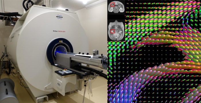
A recent study uses machine learning techniques to optimize the algorithms used to map brain connections using function MRI scans (fMRI). This is perhaps another example of how neuroscience and computer science are increasingly supporting each other. The researchers were using marmoset brains as their subject. First they mapped regions of the marmoset brain using a standard technique that involved injecting a fluorescent tracer into neurons then following where that tracer goes, through neuronal connections to other parts of the brain. This is perhaps the gold standard technique, but involves sacrificing the animal so that the brain can be sliced and examined microscopically. This technique, therefore, cannot be used in humans.
They then mapped the same regions of the marmoset brain using the less invasive technique of fMRI scanning, which can trace the movement of water through neuronal connections in order to map them. The fMRI method, however, is not nearly as precise as the fluorescent tracer technique. It also suffers from many trade-offs, which the researcher likened to camera settings. You can change the settings to produce very different looking pictures. The question is – which settings (which in regard to the fMRI refers to the computer algorithms that analyze the data) give the best picture of the connectome?
In order to determine this they compared the fMRI results to the fluorescent tracer results. They then used an evolutionary algorithm to iterate the fMRI connectome algorithm until it produced the best results. The evolutionary approach means that you essentially choose multiple sets of random parameters (although within a range known to be functional) and then pick the set that produced the best results (compared to the fluorescent tracer standard), iterate the winner and run the competition again, and then keep doing this until you have optimized the algorithm.
They then tested their optimized algorithms on fresh marmoset brains (meaning, not used in the study) to see how they perform, and they did much better than the algorithms that were not optimized by this machine learning technique. They did note, however, that even with this optimization, there are still trade-offs. Specifically, algorithms could be optimized for the long distance neurons, or for better images for short range neurons, but not simultaneously for both. However, this just means that they will need to run various optimized algorithms over the same region to get both short and long range neurons (like taking multiple pictures with different settings and combining them into one picture with high dynamic range).
The benefit of this research is that neuroscientists can use a non-invasive technique to map human brains with higher fidelity (fewer false positive and false negative connections). The authors also caution about using non-optimized algorithms, because their research also shows how unreliable this technique can be. Hopefully the machine learning technique can be further improved, increasing the optimization of the fMRI brain mapping technique.
Mapping the connectome, and understanding in greater detail how brain networks function, may in turn help advance neural network computing, which is designed to mimic the function of the brain. Reproducing brain networks in computers will help us better understand how brain networks function. The ultimate expression of this self-reinforcing process may be an entire human brain mapped in silico, but not just mapped, made into a functional neural network computer. This will massively accelerate research into how neural networks work, including what happens in various disease states.
The question is – will a functioning complete map of the human brain connectome be, itself, a conscious self-aware entity? I think the answer, almost by definition, is yes. If it is a full map of the human connectome and the connections are functional, using something like neural networks that function like actual neurons, then it would have to have all the functions of a human brain, including consciousness. In fact, I suspect this will be the actual path we take to producing the first general AI. That would complicate further research, however, because it would introduce ethical considerations. If the artificial brain is self-aware, we should not just experiment on it heedless of its experience and what rights it is entitled to.
But perhaps one of the first things we will discover is how to keep such an entity from being conscious, so that we can conduct research on its subsystems without ethical considerations. In fact, that will already be happening long before we make a fully conscious neural network. We are already, for example, conducting research on our connectome models of the visual system.
It seems clear, however, that neuroscience and computer science will increasingly benefit from advances in the other discipline, and to some extent they will merge.