Apr 20 2021
Imaging the Living Brain In Action
 One major factor in the progress of our understanding of how brains function is the ability to image the anatomy and function of the brain in greater detail. At first our examination of the brain was at the gross anatomy level – looking at structures with the naked eye. With this approach we were able to divide the brain in to different areas that were involved with different tasks. But it soon became clear that the organization and function of the brain was far more complex than gross examination could reveal. The advent of microscopes and staining techniques allowed us to examine the microscopic anatomy of the brain, and see the different cell types, their organization into layers, and how they network together. This gave us a much more detailed map of the anatomy of the brain, and from examining diseased or damaged brains we could infer what most of the identifiable structures in the brain did.
But still, we were a couple of layers removed from the true level of complexity of brain functioning. Electroencephalography gave us the ability not to look at brain anatomy but function – we could detect the electrical activity of the brain with a series of electrodes in real time. This gave us good temporal resolution of function, and a good window into overall brain function (is the brain awake, asleep, or damaged) but very poor spatial resolution. This has improved in recent decades thanks to computer analysis of EEG signals, which can map brain function in higher detail, but is still very limited.
CT scans and later MRI scans allow us to image brain anatomy, even deep anatomy, in living creatures. In addition we can see some pathological details like edema, bleeding, scar tissue, iron deposition, or inflammation. With detailed imaging we could see the lesion while still being able to examine a living patient (rather than having to wait until autopsy to see the lesion). As MRI scans advanced we could also correlate non-pathological anatomical features with neurological function (such as skills or personality), giving us yet another window into brain function.
Function MRI scanning (fMRI) and Positron Emission Tomography (PET) allow us to image brain function in real time in living creatures, mostly by inferring brain activity through metabolic markers. With increasingly high resolution we could see which parts of the brain “light up” during specific cognitive states or activities. The temporal and spatial resolution of these techniques, however, remains limited. Further, subjects need to be still in a scanner, which limits the conditions in which they can be scanned.
The ultimate goal is to create a scanning method that can image brain anatomy and function at a single-neuron level of spatial resolution and with high temporal resolution (think of this as frames per second), and that will cover the entire brain, in a portable device that a living creature can wear or have implanted without interfering with their normal activity. (That’s not too much to ask for, is it?) We may achieve this by building an exact replica of the brain in a computer, either virtually or in a silicon brain, but in order to get there we may need to be able to scan a living brain first. We are probably decades away from having anything close to this technology, but we are making progress in getting part of the way there.
The closest technology we have now is optogenetics. With this technology we genetically modify neurons to either be activated by specific frequencies of light or to fluoresce in response to light. The advantage of this technique is that is has the potential for single neuron resolution. The main limiting factor is the ability to miniaturize the laser to generate the light and the optics to record the signal. There are also different techniques used with different advantages and disadvantages, in terms of resolution and image quality. The lack of adequate miniaturization practically limit the technique to transparent tissue, either transparent organisms or tissue samples that have been chemically cleared.
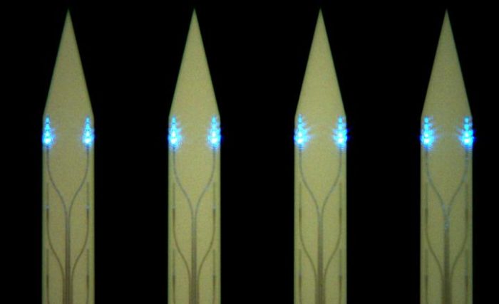
A new study reports a significant advance in the technology use for one technique called light sheet fluorescence brain imaging. With this technique a laser illuminates a thin slice of tissue (as opposed to other techniques which illuminate a point) perpendicular to the device that images the sample. This produces good spatial and temporal resolution, partly because it is much faster than single point techniques. But again, the main limiting factor in terms of reaching our ultimate goal is miniaturizing the laser and lens. The advance being reported is the creation and testing of a miniature light-sheet generator, or a photonic neural probe, that is small enough to be transplanted in a living brain. This also means the technique could be applied to non-transparent organisms, such as a mouse.
These tiny probes will be a useful advance for light sheet fluorescence imaging, and gets us one step closer to our goal, but we sill need to further miniaturize the recording optics sufficiently that a test animal could be free moving with the scanner implanted. So again, we are not there yet, but this is a nice step in the right direction.
While our perfect goal may be decades away, we are starting to finally get down to single-neuron level of brain functional imaging. This technology will help us achieve another goal, mapping the human “connectome” – all the circuits that make up a human brain. This would be an amazing achieving, on the level or perhaps even greater than mapping the genome.