
Oct 07 2021
Map of the Primary Motor Cortex Published
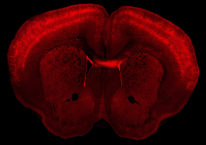 By now, especially if you are a regular reader here, you have probably heard of the connectome project, an attempt to entirely map the cells and connections of the human brain. This goal is actually comprised of multiple initiatives, one of which is the Brain Research Through Advancing Innovative Neurotechnologies (BRAIN) funded by the NIH. They have now published in Nature their first major result – a map of the mammalian primary motor cortex (technically a “multimodal cell census and atlas of the mammalian primary motor cortex”).
The goal of this initiative is to break the brain down into its constituent parts and then see how they all fit together. This begins with knowing all the different brain cell types, and this is part of the string of publications they have produced. The brain contains about 160 billion cells, with 87 billion neurons and the rest astrocytes (which provide supporting and modulating functions). There are many different kinds of neurons, with significant functional differences. Neurons differ in their structure and their chemistry.
The basic structure of a neuron is a cell body with dendrites (hair-like projections) for incoming signals and axons (longer projections) for outgoing signals. But the shape, number, and arrangement of dendrites and axons can vary considerably, and reflect their function, which relates to the pattern of connections the neuron makes. Neurons also differ in terms of their biochemistry – which neurotransmitters do they make, and which neurontransmitter receptors they have. Some neurotransmitters like glutamate are activating (make neurons fire faster) and others like GABA are inhibitory (make them fire slower or not at all).
The new studies detail 116 different cell types in the motor cortex. That is more than the previous list, but many of these new cell types are subtypes or variations on known neuronal types. So the research was more about getting to a finer level of detail, rather than discovering new and previously entirely unknown neurons. Part of the complexity of the brain is that cell types, neurotransmitters, and receptors are not divided into clean types. This is because the brain evolved, and evolution is messy. There are variations on variations, with each neurotransmitter having multiple versions, with different affinities for different receptor subtypes and complex distributions within different brain areas and for different circuits.
The researchers used various methods to characterize the different neuronal types:
The scRNA-seq technique was one of nearly a dozen separate experimental methods used by the BICCN team to characterize the different cell types in three different mammals: mice, marmosets and humans. Four of these involved different ways of identifying gene expression levels and determining the genome’s chromatin architecture and DNA methylation status, which is called the epigenome. Other techniques included classical electrophysiological patch clamp recordings to distinguish cells by how they fire action potentials, categorizing cells by shape, determining their connectivity, and looking at where the cells are spatially located within the brain. Several of these used machine learning or artificial intelligence to distinguish cell types.
They looked at gene expression to see what which neurotransmitters and receptors it was producing. They also looked at how the neurons behaved electrically in response to different stimuli – what happens when we put some GABA on this neuron? They also mapped the connections those types of neurons make within the motor cortex.
The motor cortex is probably a good place to start for mapping the brain. Although we are bound to find hidden complexity (that’s kinda the point) the function and anatomy of the motor cortex is already well understood. Neurons in the motor cortex activate when we want to move, sending signals through a single axon down deep into the brain, through the brain stem, then to the spinal cord where the axon synapses on the anterior horn cell. That neuron then sends a second axon to the group of muscles to be contracted. The signal from cortex to muscle goes through only two neurons, containing the longest axons in the body.
There is, of course, a lot more to it than that. There is the premotor cortex which is involved in planning and executing voluntary movement and ultimately activates the motor neurons. About a third of the motor neurons do not connect directly to the spinal cord, but instead go to the cerebellum, which is involved in coordination. There various motor cortex signals are processed so that movements can be coordinated together. The cerebellum allows for balance, precision, and timing. The cerebellum then modulates motor cortex signals to the muscles. There is also the basal ganglia which calibrates the strength of the signals connecting motor planning to motor execution. This way you move as much as you want to, not more or less. There are also multiple types of sensory feedback, a lot of which is also processed in the cerebellum, so that we can adjust our movements to what we see, our sense of where our body parts are and the state of contraction of our muscles, and our vestibular sense of gravity and movement.
That’s a basic schematic of voluntary motor movement, but the goal of the BRAIN initiative is to map all these connections and cell types in complete detail. Even mapping one system in the brain, like the motor system, is just getting started. Next they will want to completely map the sensory system, visual, hearing, and taste. We can then get to some of the more complex areas of the brain, like language function, calculations, and visuo-spatial reasoning. The frontal lobes, which contain executive function and higher level reasoning, will likely be the most complex. We also need to map how this all connects together.
Connectome projects like this also feed into other projects that are attempting to model the brain in a computer simulation. These two lines of research work well together, with each feeding into the other. Of course the most interesting question is – what happens when we have a complete map of the human brain simulated in a supercomputer, and we turn it on?
Beyond computer simulations, this research is likely to have far-reaching implications for neuroscience. I won’t bother to speculate about specific applications. I always find that gratuitous. This is basic science, expanding our understanding of how the brain works. Potential applications are many, some obvious and others likely to be surprising. For now, understanding the connections of the brain for its own sake is enough.