Jul 04 2008
The Not-So-Intelligent-Design of the Human Eye – Part II
Yesterday I discussed the difference between the bottom-up design of evolution vs the supposed top-down design of a god/intelligent designer. I then used the human eye as an example of bottom-up design, made manifest by quirky and sub-optimal aspects that do not make sense from a top-down perspective. Today I will give further examples of the sub-optimal design of the human eye which predisposes to certain opthalmological diseases and disorders.
Angle Closure Glaucoma
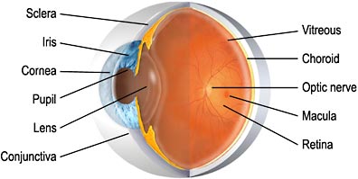
Glaucoma results from increased pressure inside the eye. Between the iris and the cornea is a space called the anterior chamber which is filled with the aqueous humor – a clear liquid that is constantly made and drained from this chamber. If the drainage is blocked or slowed then the fluid will back up, increasing the pressure inside the eye. This pressure can damage the optic nerve resulting in blindness. In acute angle closure glaucoma blindness can occur within hours and it is therefore an emergency.
The draining channels, called the trabecular meshwork, are at the angle between the iris and the cornea. When this angle gets too narrow, because the iris is pushing up against the meshwork, then drainage can be blocked. This is another example of sub-optimal design – the drainage is unnecessarily precarious and susceptible to blockage, leading to rapid loss of vision.
Through the quirkiness of genetics, some populations are more susceptible to angle closure glaucoma than others because they have an inherently narrower angle to start with. The most susceptible populations are Asians and Inuit.
Macular Degeneration
The macula is that part of the retina that has the most dense concentration of rods and cones for detailed vision. Within the macula is a smaller area called the fovea which contains only cones and has the highest density of these receptors. The very existence of the macula, however, is a partial fix for the problem I discussed in Part I – that the retinal layers are “backwards” with the nerve and blood vessels between the receptors and the direction of light. This limits the density of rods and cones, and so the partial fix is to have one small area cleared of nerves and blood vessels where rods and cones can be more dense. However, if the human retina were designed like that of the squid and other cephalopods this would not be necessary.
The dependence of the human eye on the macular for sharp vision creates a vulnerability, for any problem with that small area will have a dramatic effect of visual acuity. The rest of the retina will not be able to adequately compensate for the loss or compromise of the macula because the density of rods and cones are just too diffuse.
Macular degeneration is the most common cause of blindness. Although the cause is often unknown its severe effects on vision are a consequence of the need for a macula as a partial fix for the poor retinal design.
Detached Retina
A yet another consequence of the “backward” arrangement of the retina is the susceptibility to detached retina. This occurs when the photoreceptor layer becomes detached from the pigment epithelium beneath. The subsequent loss of nourishment causes blindness in the detached part of the retina. One cause of retinal detachment is minor trauma to the retina that allows fluid to leak and build up between the retina and the pigment epithelium, thus separating the layers. But the more common cause is simple aging that results in subtle changes to the shape of the globe of the eye and loss of the elasticity of the vitreous humor – the gel that fills the globe.
Again, the cephalopod eye does not suffer from retinal detachment because the axons from the photoreceptors anchor them to the layers beneath.
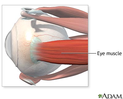
The Extraocular Muscles
The arrangement of the extraocular muscles – the muscles that move the eyes, is also difficult to explain from a top-down engineering perspective. There are more muscles than are minimally necessary and yet there is no functional redundancy. In order to move a sphere in any direction only three muscles would be necessary, evenly spaced like the legs of a tripod. The human eye has six – the superior, inferior, lateral, and medial rectus, and the superior and inferior oblique. And yet, despite the extra three muscles, the loss of function of any one muscle causes an impairment of eye movement and results in double vision or displaced vision. A more frugal design with only three muscles would be more efficient and less prone to malfunction, as there are fewer components to break down.
If the eye were to be designed with more than the minimal three muscles, then it would make sense to arrange the muscles so that the loss of one or even more would not impair eye movement.
The configuration of cranial nerve control of the extraocular muscles also makes no design sense. The lateral rectus is controlled by cranial nerve VI (abducens), the superior oblique by cranial nerve IV (trochlear) and the rest by cranial nerve III (oculomotor). There is no functional advantage to this particular arrangement; it is an accident of evolution. Having three cranial nerves responsible for eye muscles multiplies the opportunity for failure of any one, and again there is no redundancy as a hedge against malfunction.
But the most suboptimal aspect of cranial nerve innervation of the extraocular muscles is the abducens, cranial nerve VI. The abducens takes an unnecessarily long path from the brainstem, through the skull, and to the lateral rectus. There is no design reason for this long path – it too is an accident of evolution. It makes the abducens particularly vulnerable to injury or stretching, and for this reason abducens palsy is the most common extraocular muscle weakness. The trochlear nerve (cranial nerve IV to the superior oblique) also takes an unusual pathway – it exits the brainstem heading toward the back of the brain, which is the wrong direction. It must then swing around and head toward the eyes. As with the abducens nerve, this unnecessarily long pathway increases the potential for malfunction.
Susceptibility to disease
The human eye is susceptible to a number of other diseases and dysfunctions that do not result from any obvious “design flaw” but for which there is no particular protection either. This is a weaker line of evidence for evolution, because there is no clear flaw to point to, simply the lack of design elements that could have protected the eye from problems. For example, if the shape of the eye and cornea are not within certain narrow parameters then the image will not focus on the retina, leading to near-sightedness or far-sightedness. It is not difficult to imagine plausible mechanisms to correct these so-called refractive errors – such as mechanisms for distorting the shape of the cornea or the globe of the eye. This could be done with muscles or by increasing or decreasing the fluid behind the cornea and within the globe.
Cataracts form when the proteins in the lens begin to clump together, causing the lens to become cloudy and obscuring vision. A protein design that does not allow for this clumping, or a repair mechanism to replace clumped proteins, could render the lens immune to cataracts. The lens also tends to become stiff with age, reducing its ability to focus near objects onto the retina. As a result the near point – the closest point that a person can bring into focus, moves slowly farther away, causing what is called presbyopia. Most people, when they get into their 40’s or 50’s, need to use reading glasses for this reason.
Conclusion
Biologist Frank Zindler said, “As an organ developed via the opportunistic twists and turns of evolutionary processes, the human eye is explainable. As an organ designed and created by an infinitely wise deity, the human eye is inexcusable.” Any objective view of the human eye shows this to be clearly true.